Table of Contents
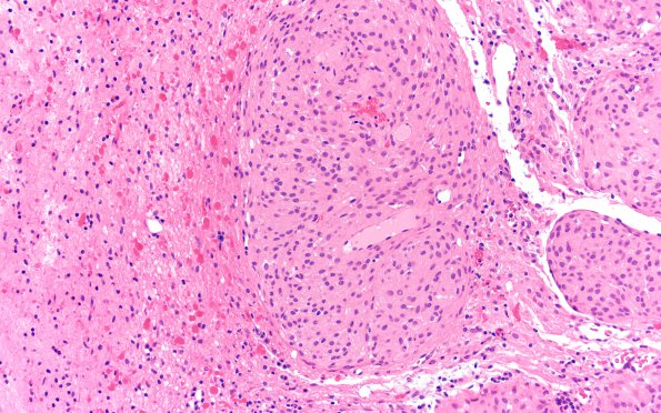
Washington University Experience | NEOPLASMS (MENINGIOMA) | Brain Invasion | 9A3 Meningioma, brain invasion (Case 9) H&E 20X
H&E stained tissue shows a neoplasm arranged in lobules. The cells have a syncytial pattern with indistinct cell borders and abundant eosinophilic cytoplasm. No mitoses are identified. Psammoma bodies are present. Brain invasion is identified with areas of reactive Rosenthal fibers.

