Table of Contents
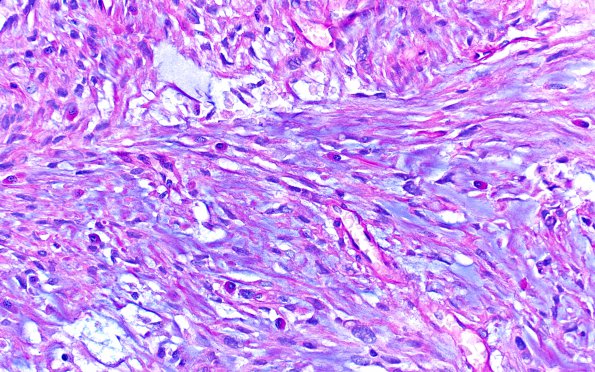
Washington University Experience | NEOPLASMS (MENINGIOMA) | Chordoid | 10A1 Meningioma, Chordoid (Case 10) Alcian Blue 1
Case 10 History ---- The patient was a 30-year-old woman with history of recent changes in papilledema. MRI showed a 5.4 cm enhancing dural based circumscribed mass involving the left frontoparietal lobe, most consistent with meningioma. Operative procedure: Left-sided craniectomy for tumor resection. ---- 10A1-2Hematoxylin and eosin-stained sections demonstrate a neoplasm characterized by meningothelial cells set in a predominantly myxoid background, arranged variably in small clusters, cords and trabecular architecture which occasionally form whorls. A less predominant area appears fibroblastic. Most of the cells are characterized by eosinophilic cytoplasm, ovoid nuclei with relatively evenly dispersed chromatin, frequent intranuclear cytoplasmic inclusions and nuclear clearing, and scattered hyperchromatic forms. There is increased cellularity and focal sheeted architecture; however, other atypical features including small cell change, necrosis and prominent nucleoli are not seen. Mitotic figures are scattered and, focally reaches up to 4/10 HPF. In some areas, the vessels appear dilated and congested. There is a thin rim of brain parenchyma, focally suspicious of brain invasion. The tumor also invades the dura. Ancillary studies were performed on block A4. Alcian blue/PAS special stain highlights Alcian blue positive predominant myxoid stroma. GFAP confirms focal brain invasion. A Ki-67 demonstrates a proliferation index which reaches ~7.6% in most active foci.

