Table of Contents
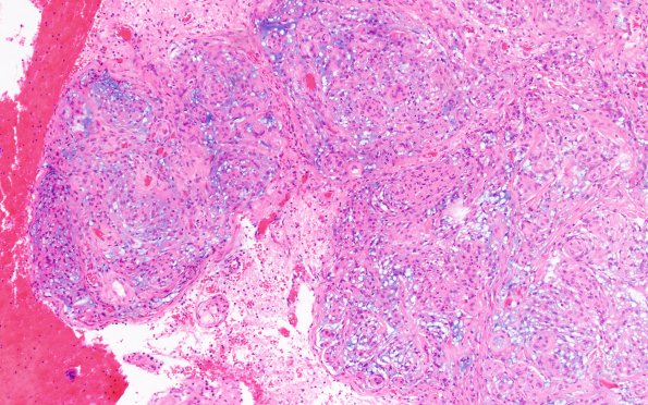
Washington University Experience | NEOPLASMS (MENINGIOMA) | Chordoid | 11A1 Meningioma, chordoid & invasion (Case 11) H&E 4X
Case 11 History ---- The patient was a 40-year-old woman presenting with headache with imaging showing a 5 cm enhancing extra-axial right occipital lesion. Operative procedure: Right craniotomy for resection of tumor. ---- 11A1-4 H&E shows a moderately cellular neoplasm composed of syncytial cells in nests and whorls. Mitoses are rare (1/10HPF). There are focal myxoid/chordoid changes in the stroma as well as multifocal areas of necrosis. No additional atypical features are present. Brain invasion is identified.

