Table of Contents
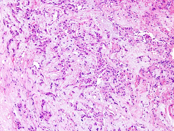
Washington University Experience | NEOPLASMS (MENINGIOMA) | Chordoid | 15A Meningioma, chordoid (Case 15) H&E 6.jpg
Case 15 ---- The patient was a 50 year old woman with a history of meningioma with chordoid features in the right sphenoid wing and periorbital area, six years prior. Follow-up imaging shows recurrent tumor. Operative procedure: Right craniotomy for resection of tumor, lumbar drain placement and orbital reconstruction. ---- 15A. A significant subset of the tumor cells shows chordoid features represented by strands and cords of eosinophilic cells with vacuolated cytoplasm, situated in a myxoid stroma. No necrosis, small cell change, or macronucleoli are identified. Mitotic figures are inconspicuous. The tumor cells are organized in small nests and strands, as well as singly scattered cells in an extensively myxoid background. Mitotic figures are inconspicuous. No necrosis, small cell change, or macronucleoli are identified. ---- We compared the current tumor with the patient's prior excision specimen 6 years prior and the current tumor shows predominance of chordoid meningioma which comprised only a small subset of tumor cells in the prior specimen.

