Table of Contents
Washington University Experience | NEOPLASMS (MENINGIOMA) | Chordoid | 2A1 Meningioma, Choroid (Case 2) d
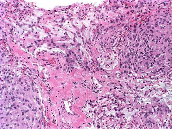
Case 2 History ---- The patient was a 67 year old woman with schizophrenia and a dura-based enhancing mass. She presented with a bleed in the tumor mass. Operative procedure: Craniotomy and resection. ---- 2A1-4 This is a dural based mass composed of epithelioid cells with uniform round to oval nuclei, occasional macronucleoli, and moderate amounts of clear to eosinophilic cytoplasm arranged in sheets or whorls. In areas, the tumor cells show a chordoid-like or trabecular pattern in a myxoid background. There is extensive hemorrhage and the mitotic count reaches 6/10HPF. The histology is consistent with that of an atypical meningioma with clear cell and chordoid features (WHO Grade 2). The clear cell component shows cytoplasmic positivity for PAS and is negative with diastase. Immunohistochemical stains performed show that the tumor cells have nuclear positivity for progesterone receptor in the chordoid area and rare cells are positive in the solid/clear cell component. The Ki67 labeling index is high in the solid component.

