Table of Contents
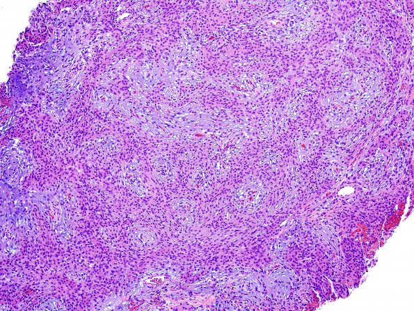
Washington University Experience | NEOPLASMS (MENINGIOMA) | Chordoid | 5A1 Meningioma, Chordoid (Case 5)
Case 5 History ---- The patient was a 70 year old man with a history of prostate cancer, who experienced abrupt onset mental status change. Radiologic imaging revealed bifrontal intracerebral, subarachnoid and intraventricular hemorrhage, and an extra-axial, T1 isointense, T2/FLAIR hyperintense, homogeneously enhancing, anterior fossa skull base mass along the cribriform plate and planum sphenoidale, which measures 3.7 x 2.67 x 2.65 cm. Operative procedure: Right frontal craniofacial resection and zygomatic osteotomy. ---- 5A1-3 Histological sections show a dura associated neoplasm with two predominant and intermixed histologic patterns. The majority of the tumor tissue exhibits a variably pale to intense basophilic myxoid or mucinous background, in which eosinophilic spindled or stellate tumor cells appear, occasionally forming tenuous cords, reminiscent of chordoma. Intermixed with this 'chordoid' pattern is the other, which is formed by similar tumor cells arranged in fascicles and whorls. The tumor cells in both phases have round-to oval nuclei, relatively open, speckled chromatin, small nucleoli, occasional intranuclear pseudoinclusions, eosinophilic cytoplasm, and indistinct cell borders, wherever cell-cell contacts occur. Rare microcalcifications are present. Several focal areas of hemosiderin deposition are noted, consistent with previous hemorrhage. Mitotic figures are rare (<1/10 HPF). There is no evidence of hypercellularity, loss of histological architectural pattern ('sheeting'), macronucleoli, increased nucleus-to-cytoplasm ratio ('small cell change'), or necrosis. No brain parenchyma is identified to allow assessment of brain invasion. (H&E)

