Table of Contents
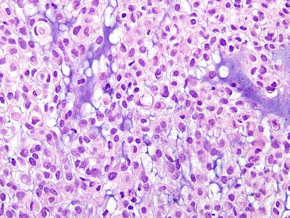
Washington University Experience | NEOPLASMS (MENINGIOMA) | Chordoid | 6B2 Meningioma, chordoid (Case 6) H&E 8.jpg
A a region comprising much less than half of the sampled tissue shows clear cell changes, myxomatous material is seen between tumor cells giving these regions a chordoid appearance. Frequent mitoses are seen with up to 4/10HPF. Immunostaining for Ki67 demonstrates a proliferation index of 18%.

