Table of Contents
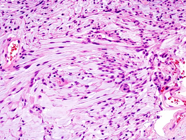
Washington University Experience | NEOPLASMS (MENINGIOMA) | Chordoid | 7C3 Meningioma, Chordoid (Case 7) H&E 3
Tumor cells have abundant eosinophilic cytoplasm, vesicular nuclei focally containing intranuclear inclusions. In some areas, the tumor cells are arranged in a cord-like pattern in a myxoid background (chordoid areas). However, the chordoid areas represent a relatively small percentage of the total tumor (~15-20%). Mitoses are rare and necrosis, hypercellularity or small cells are not evident. ---- Ancillary data (not shown): Immunohistochemically, the tumor cells are diffusely positive for progesterone receptor and show patchy positivity with epithelial membrane antigen. The tumor has a low proliferation rate (Ki-67 labeling index ~ 2.7%). A similar immunoprofile is noted within chordoid areas as well. The overall morphological and immunohistochemical findings are consistent with a meningioma with focal chordoid areas, WHO grade I. Given the relatively small percentage of the tumor that shows chordoid features, at that time we felt that this tumor did not merit a designation of WHO grade 2 but I think that it probably merits that diagnosis now.

