Table of Contents
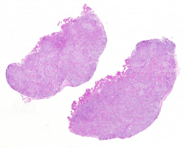
Washington University Experience | NEOPLASMS (MENINGIOMA) | Chordoid | 8B1 Meningioma, Chordoid (Case 8) H&E WM
8B1-5 Sections of intradural tumor reveal a vaguely lobulated moderately cellular tumor composed of cords of small eosinophilic, often vacuolated epithelioid cells, set within an abundant matrix of basophilic mucin, with lobules demarcated by thick fibrous septae. These chordoma-like areas are mixed with foci of more classic meningioma, the latter shown the typical whorls of large epithelioid to spindled cells. Mitotic figures are hard to find and there are no areas of tumor necrosis, small cell formation, hypercellularity, or macronucleoli.

