Table of Contents
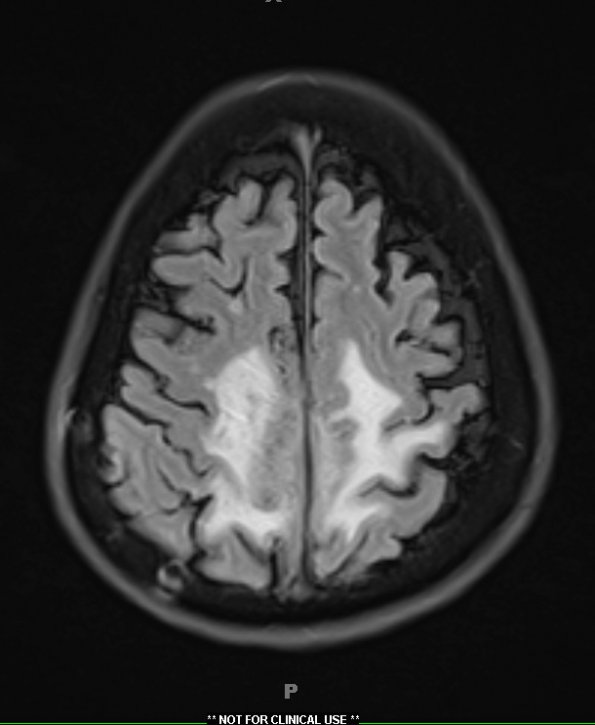
Washington University Experience | NEOPLASMS (MENINGIOMA) | Chordoid | 9A1 Meningioma, chordoid (Case 9) TIRM 1 - Copy
Case 9 History ---- The patient was a 58-year-old woman with a history of Moya-moya disease (diagnosed 6 years previously) and a large frontoparietal parafalcine meningioma, resected more than 20 years previously at an outside institution. Later review of that material at WUSOM found a fibrous meningioma, WHO grade. In 2003, she underwent gamma knife radiosurgery for residual/recurrent tumor associated with the superior sagittal sinus on the right. Recently (2016), she developed significant paraparesis and difficulty with the use of her right hand and imaging demonstrated tumor recurrence. Operative procedure: Craniotomy for tumor resection with intraoperative MRI. ---- 9A1,2 MRI studies showed a frontoparietal parafalcine mass with TIRM (9A1) and T1-weighted, contrast applied (9A2) hyperintensity in the left parafalcine region. There were also fingerlike projections from the left parafalcine component of the mass consistent with brain invasion, and interval growth of an enhancing right-sided nodule.

