Table of Contents
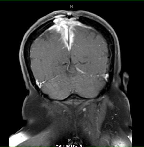
Washington University Experience | NEOPLASMS (MENINGIOMA) | Chordoid | 9A2 Meningioma, chordoid (Case 9) T1W 3 - Copy
MRI studies showed a frontoparietal parafalcine mass with T1-weighted, contrast applied hyperintensity in the left parafalcine region. There were also fingerlike projections from the left parafalcine component of the mass consistent with brain invasion, and interval growth of an enhancing right-sided nodule.

