Table of Contents
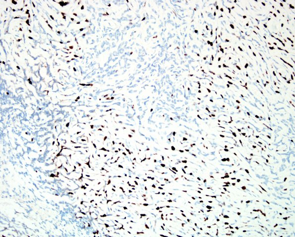
Washington University Experience | NEOPLASMS (MENINGIOMA) | Chordoid | 9C Meningioma, chordoid (Case 9) Ki67 1.jpg
IHC for proliferation marker Ki-67 stains a large subset of tumor nuclei in a diffuse pattern, ranging up to at least 26% in hypercellular areas with higher mitotic indices. ---- Ancillary data (not shown): Immunohistochemistry for GFAP stains adherent brain parenchyma, highlighting areas of brain invasion. IHC for progesterone receptor is negative. Residual/recurrent chordoid meningioma with brain invasion and focal anaplasia, WHO grade 3.

