Table of Contents
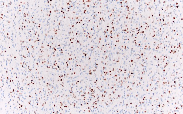
Washington University Experience | NEOPLASMS (MENINGIOMA) | Meningioma - Clear cell | 12A Clear cell meningioma (Case 12) Ki67 20X
Case 12 History ---- The patient was a 69 year old woman with a prior history of renal cell carcinoma. She underwent craniotomy for a right frontal mass in 2005 that recurred in 2006. ---- Specimens from the 2005 resection specimen reveal a meningothelial neoplasm which appears well differentiated focally, including well-formed whorls, small rounded nucleoli with scattered nuclear pseudoinclusions and abundant eosinophilic cytoplasm. In other areas, the tumor is predominantly arranged in sheets of clear cells interrupted by densely eosinophilic perivascular and interstitial collagen bundles. Collections of small cells with decreased cytoplasm are seen focally. In some areas, there is increased nuclear prominence and increased cellularity. The mitotic index is elevated, focally reaching 14 mitoses per 10 high power fields. Additionally, brain invasion is seen focally. The diagnosis of the 2005 specimen is atypical meningioma with clear cell features, WHO Grade 2.---- 12A Tumor cells in the 2005 specimen show EMA reactivity (EMA IHC)

