Table of Contents
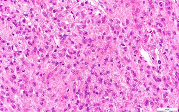
Washington University Experience | NEOPLASMS (MENINGIOMA) | Meningioma - Clear cell | 12C3 Clear cell meningioma (Case 12) H&E 40X
12C3 Note the large numbers of mitoses in the 2006 specimen, focally reaching up to 44 mitoses per 10 high power fields (H&E) ---- Ancillary results (not shown): Both specimens show rich intracytoplasmic glycogen deposition with strong and nearly diffuse PAS positivity that disappeared after diastase digestion. The MIB-1 (Ki-67) labeling index was 16.6% for the 2005 specimen and 38% for the 2006 specimen. The tumor cells in the 2006 specimen are negative for epithelial membrane antigen and PR immunoreactivity. ---- The morphologic, histochemical, and immunohistochemical features are consistent with the diagnoses of atypical meningioma with clear cell features in the 2005 specimen and anaplastic meningioma with clear cell features in the 2006 specimen. ---- Additionally, fluorescence in situ hybridization (FISH) was performed on the 2006 specimen utilizing paired probes against CEP9 and p16 (9p21). A deletion of the p16 region was found in this particular case, consistent with the diagnosis of anaplastic meningioma in general and the more aggressive subset in particular. The diagnosis made was anaplastic meningioma with clear cell features, WHO Grade 3, clinically recurrent

