Table of Contents
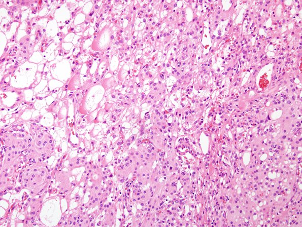
Washington University Experience | NEOPLASMS (MENINGIOMA) | Meningioma - Clear cell | 13A1 Meningioma, atypical, microcystic-clear cell (Case 13) H&E 1
Case 13 History ---- The patient was a 62 year old woman who presented with symptoms of a stroke who was found on MRI to have an enlarging, extra-axial, dural based, (non-uniform) contrast enhancing left frontal lobe mass. Operative procedure: Craniotomy with tumor resection.---- 13A1-4 The tumor cells are large and elongated with eosinophilic cytoplasm, many with a more epithelioid shape and distinct cell borders, some with cleared cytoplasm and peripherally located nuclei. In some regions numerous blood vessels, ranging from large thin walled structures to small capillaries, give the tumor an angiomatous appearance. While in most areas there is a whorled confirmation of the tumor cells and some sheeted architecture is noted. Small cell change, hypercellularity, and spontaneous necrosis are not identified. Occasional examples of nuclear pseudoinclusions are found. Mitotic activity is frequent with up to 5/10HPF.

