Table of Contents
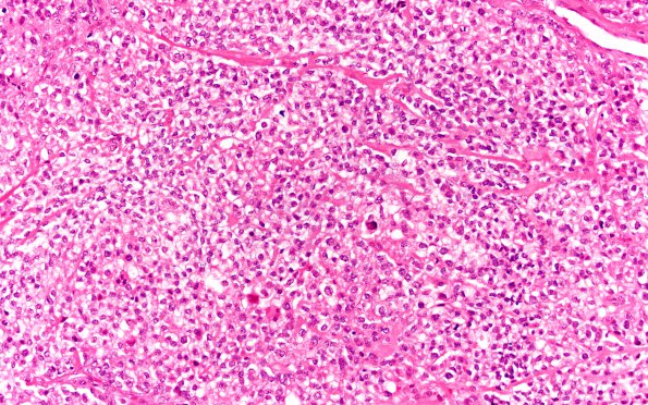
Washington University Experience | NEOPLASMS (MENINGIOMA) | Meningioma - Clear cell | 21A1 Clear cell meningioma (Case 21) H&E 20X
Case 21 History ---- The patient was a 54 year old woman who presented with headache and hypertension. Imaging revealed extensive meningeal enhancement over the left hemisphere, associated with marked mass effect and midline shift. The diagnosis of leptomeningeal carcinomatosis was favored. ---- 21A1,2 This is a markedly cellular epithelioid neoplasm arranged primarily in sheets and vague lobules. There is moderate nuclear pleomorphism and the tumor cells have a moderate quantity of predominantly clear cytoplasm. The tumor nuclei are round to oval, with occasional bizarre or multinucleate forms identified. Focal brain invasion is evident. The mitotic index is brisk, focally reaching up to 25 mitoses/10HPF. Foci of necrosis are evident.

