Table of Contents
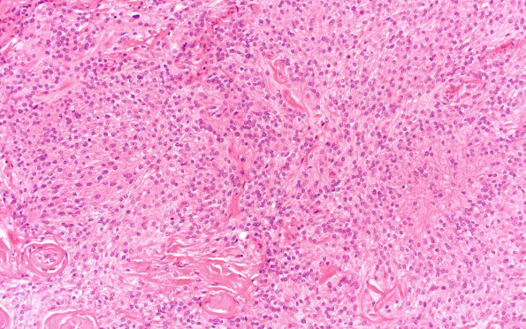
Washington University Experience | NEOPLASMS (MENINGIOMA) | Meningioma - Clear cell | 22A1 Meningioma, clear cell (Case 22) H&E 20X
Case 22 History ---- The patient was a 66 year old woman with a clinical diagnosis of meningioma. She presented in October 2003 with some uncontrollable movements of the left hand, associated with weakness which lasted for an hour. MRI at outside hospital revealed a 3 x 4 cm lesion of the right convexity. Operative procedure: Right craniotomy and tumor resection. ---- 22A1,2 Sections showed a hypercellular neoplasm composed of spindled to epithelioid cells with round to oval, uniform, vesicular nuclei and moderate amounts of eosinophilic to amphophilic cytoplasm forming sheets with scattered whorls. There is also blocky perivascular collagen deposition in these areas consistent with focal clear cell histology. There are areas with small cell change and scattered cells with large, pleomorphic nuclei. Some of the cells focally have macronucleoli. Occasional mitoses are seen with a mitotic count of 2-3/10HPF.

