Table of Contents
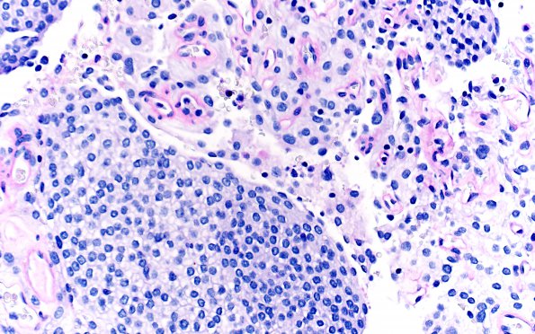
Washington University Experience | NEOPLASMS (MENINGIOMA) | Meningioma - Clear cell | 23B3 Clear cell meningioma (Case 23) PAS-D 40X 2
PAS-positive cytoplasm of tumor cells is described in the request for consult submitting report. Those slides are no longer available. The images shown are those of PAS with diastase treatment. The latter is consistent with glycogen accumulation as the explanation for the clear cell morphology. Tumor cells have no evidence of PAS staining but vessel walls do, a pattern which is consistent with glycogen deposition in a clear cell meningioma. ---- Ancillary data (not shown): There is focal EMA positivity. The Ki-67 labeling index is low. Additional stains were performed at Washington University on submitted paraffin block material. A stain for progesterone receptor displays focal strong positivity, although the majority of tumor cells are not immunoreactive. ---- Comment: The morphologic, histochemical, and immunohistochemical features are consistent with the diagnosis of atypical meningioma with clear cell and angiomatous features, WHO grade II. Based on this histology,

