Table of Contents
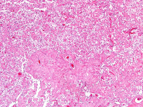
Washington University Experience | NEOPLASMS (MENINGIOMA) | Meningioma - Clear cell | 9A1 Meningioma, clear-cell (Case 9) H&E 3.jpg
Case 9 History ---- The patient was a 25-year-old woman with a history of migraine headache who presented to an outside hospital with a severe migraine associated with right face tingling, arm numbness, and nausea. MR imaging performed at BJH showed enhancing masses with central cystic spaces extending into both Meckel’s caves. The right-sided mass was contiguous with an enhancing soft tissue mass that filled the pre-pontine cistern, right cerebellopontine angle, and partially encased the basilar artery. The mass abutted the right vertebral artery, and right cranial nerves VII through X. Operative procedure: Right suboccipital craniotomy, middle fossa craniotomy, trans-mastoid approach for tumor resection. ---- 9A1-4 This is an extensively hyalinized and collagenized tumor with focal areas of sheeted cells characterized by cleared cytoplasm and polygonal shape. The nuclei of these neoplastic cells are predominantly round to ovoid, with finely granular chromatin, and occasional conspicuous nucleoli. The hyalinized components of this tumor appear to be both perivascular and interstitial and consist of coalescing blocks of collagen. Large portions of the specimen consist almost exclusively of this blocky collagen with very few nucleated cells. Mitotic activity and necrosis are not identified. Psammoma bodies are absent.

