Table of Contents
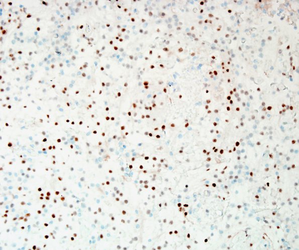
Washington University Experience | NEOPLASMS (MENINGIOMA) | Meningioma - Clear cell | 9C Meningioma, clear-cell (Case 9) PR 1.jpg
Likewise, a progesterone receptor (PR) immunostain highlights a majority of the neoplastic cells, with intensity ranging from weak to strong. ---- Ancillary Data (not shown): A Ki67 stain highlights an overall low index of proliferation (2.5%). CD34 highlights blood vessels only, not the tumor cells. CD31 highlights blood vessels, as well as a subset of tumor cells. Trying to prevent misdiagnosis of a metastatic renal cell carcinoma, the markers RCC and PAX8 were tested and were negative, results which do not support a metastatic tumor origin. A thioflavin S stain fails to demonstrate amyloid in the hyalinized areas. ---- These findings are consistent with a diagnosis of clear cell meningioma, WHO grade 2.

