Table of Contents
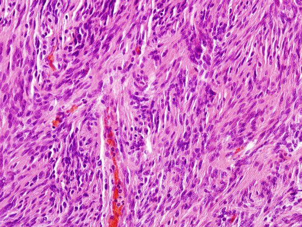
Washington University Experience | NEOPLASMS (MENINGIOMA) | Meningioma - Fibrous | 11A Meningioma, fibroblastic (Case 11) H&E 1
Case 11 History ---- The patient is a 69 year old man who experienced a fall. Radiographic imaging of the head revealed a 4.7 x 4 x 4.3 cm, homogeneously enhancing, dural based mass in the posterior fossa, displacing the right cerebellar hemisphere and fourth ventricle to the left. Operative procedure: Craniotomy. ---- 11A This is a dura-associated neoplasm composed of tumor cells with spindled morphology. The tumor cells have elongate oval nuclei, finely-speckled chromatin, small eosinophilic nucleoli, occasional nuclear clearings and pseudoinclusions, eosinophilic cytoplasm, and indistinct cell borders. Fascicles of very slender cells predominate, with only occasional whorl formation. Focally, microscopic groups of tumor cells exhibit increased nucleus-to-cytoplasm ratios ('small cell change'). Other areas show nuclear palisading, reminiscent of the Verocay bodies typical of Schwannoma. Rare areas show focal clear cell morphology. Occasional microcalcifications are present. Mitotic figures are rare, present at a density of less than 1/10HPF. There is no evidence of loss of architectural growth pattern ('sheeting'), necrosis, widespread macronucleoli, or marked hypercellularity.

