Table of Contents
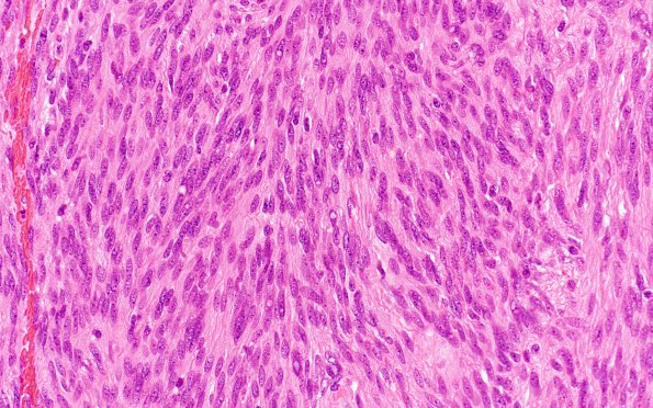
Washington University Experience | NEOPLASMS (MENINGIOMA) | Meningioma - Fibrous | 12A Meningioma, fibroblastic, atypical (Case 12) H&E
Case 12 History ---- The patient is a 67-year-old female who has a history of left parietal meningioma which was resected. Twelve years later she presented with severe headache. MRI shows a 1.5 cm enhancing dural based extra-axial right falcine mass. Operative procedure: Right frontal craniotomy for tumor resection. ---- 12A Sections show a portion of dura and a separate fragment of a meningioma where the tumor cells are arranged in whorled nests and short fascicles and have scattered prominence of nucleoli. Hypercellularity and small cell change are additionally seen. There are scattered psammoma bodies. Mitoses are easily seen, with a count of 4 mitoses/10HPF in many foci. Not shown: Immunohistochemical stains were performed that show nuclear immunoreactivity for progesterone receptor (PR) in a small subset of tumor cells with a high Ki-67 labeling index of 7.6%. ---- Comment: Overall histological features are those of meningioma. The proliferative index of 4/10HPF and presence of three of five atypical features (hypercellularity, small cell change and prominent nucleoli) are consistent with a diagnosis of meningioma, WHO grade II.

