Table of Contents
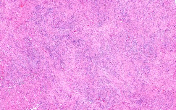
Washington University Experience | NEOPLASMS (MENINGIOMA) | Meningioma - Fibrous | 13A1 Meningioma (Case 13) H&E 4X
Case 13 History ---- The patient is a 56 year old man, who was found, on imaging studies, to have a large occipital tumor mass. MRI showed a large extra-axial, homogeneously enhancing mass in the left occipital lobe and along the left cerebellar tentorium. Clinical and radiological differential includes meningioma. Operative procedure: Stealth craniotomy for resection of brain tumor with IMRI. ---- 13A1-4 This is a neoplasm composed of spindled tumor cells organized into whorls and short fascicles. There are scattered areas of possible small cell change, however, there is no evidence of sheeting, hypercellularity, spontaneous necrosis, or brain invasion. Scattered psammoma bodies are seen. The tumor cell nuclei have finely stippled chromatin with largely inconspicuous nucleoli, although an occasional macronucleolus is noted. The mitotic rate throughout the tumor is relatively low, but focally reaches up to 2/10 HPF.

