Table of Contents
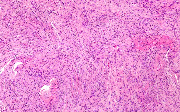
Washington University Experience | NEOPLASMS (MENINGIOMA) | Meningioma - Fibrous | 1B1 Meningioma, fibrous, intraventricular (Case 1) H&E 10X 1
1B1-4 H&E stained sections show a hypercellular, poorly circumscribed neoplasm with oval and spindled cells arranged in disordered fascicles and whorls, with wavy, elongated nuclei and inconspicuous nucleoli. Mitoses are not appreciated. Necrosis is present, notably status post radiotherapy. Occasional psammoma bodies are present.

