Table of Contents
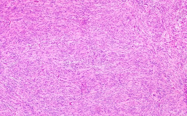
Washington University Experience | NEOPLASMS (MENINGIOMA) | Meningioma - Fibrous | 3A1 Meningioma (Case 3) H&E 10X 1
Case 3 History ---- The patient is a 58 year old woman with posterior fossa meningioma. Operative procedure: Tumor excision. ---- 3A1-3 Routine H&E stained slides of the right suboccipital tumor show fragments of tissue composed of spindled cells arranged in interlacing bundles. The tumor cells have abundant eosinophilic cytoplasm and indistinct cell borders. There is mild pleomorphism. The tumor nuclei are oval to slightly elongated, have finely stippled chromatin, and have occasional intranuclear pseudoinclusions. Psammoma bodies are also identified. Atypical features are not seen.

