Table of Contents
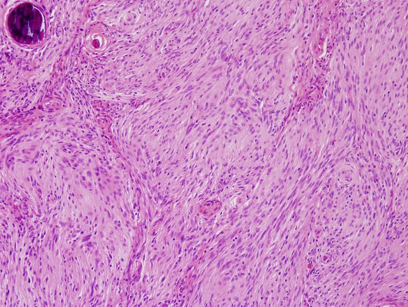
Washington University Experience | NEOPLASMS (MENINGIOMA) | Meningioma - Fibrous | 4A Meningioma (Case 4) H&E 1.jpg
Case 4 History ---- The patient is a 65 year old woman with a 3.5 x 3.4 x 3.1 cm extra-axial mass in the anterior cranial fossa adjacent to the left frontal lobe. Hypointensity within the lesion on susceptibility weighed imaging likely represents areas of calcification. Operative procedure: Stealth-guided left frontal craniotomy for tumor resection. ---- 4A Sections show a proliferation of meningothelial cells with a fibroblastic appearance arranged in broad fascicles with occasional whorls. The cells themselves are predominantly spindled with elongated ovoid to spindled nuclei, and coarsely clumped chromatin. Scattered nuclear pseudoinclusions and nuclear clearing are also seen. There are numerous psammoma bodies scattered throughout the sections. A chronic inflammatory infiltrate composed predominantly of lymphocytes is present throughout the sections. Of note, mitotic figures are rare and no subjective features of atypia, including hypercellularity, small cell change, sheeted architecture, prominent nucleoli or necrosis are identified. ---- Not shown: A PR stain shows a subset of cells with nuclear positivity. Ki-67 reveals an index of proliferation elevated to 7.2%. Overall, the histomorphological and immunohistochemical findings support a diagnosis of meningioma, WHO grade I, with elevated proliferative index.

