Table of Contents
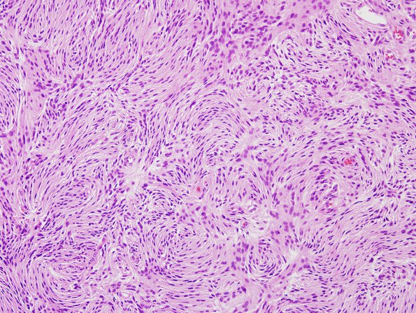
Washington University Experience | NEOPLASMS (MENINGIOMA) | Meningioma - Fibrous | 6A1 Meningioma, atypical (Case 6) H&E 2
Case 6 History ---- The patient is a 2 year old boy with a brainstem and right temporal lesion. Operative procedure: Right temporal craniotomy. ---- 6A1-3 This is a meningothelial neoplasm with a range of morphologic features. There is mild nuclear pleomorphism, with the majority of tumor cells containing round to oval nuclei and abundant eosinophilic cytoplasm. Many of the nuclei have clear holes or nuclear pseudo-inclusions. Some areas show a fibrous storiform architecture with whorls. There is vascular hyalinization. There are calcifications with psammoma bodies. Mitotic figures are easily identified, reaching 7/10HPF and there is tumor necrosis. The Ki-67 labeling index is moderate to high, reaching 11.2% focally. The morphologic features, with the presence of hypercellular areas, increased mitotic index and necrosis are different from the earlier biopsy material. The current tumor is consistent with an atypical meningioma, WHO grade II.

