Table of Contents
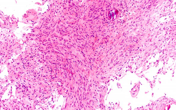
Washington University Experience | NEOPLASMS (MENINGIOMA) | Meningioma - Fibrous | 7B Meningioma, fibroblastic (Case 7) H&E 20X
7B H&E stained sections of the resected material from the intradural extramedullary, thoracic spinal cord lesion show a meningioma with a predominantly fibrous histologic pattern and scattered psammomatous calcifications. There is a modest chronic inflammatory infiltrate percolating throughout the tumor. Tumor cells show no evidence of hypercellularity, prominent nucleoli, sheeted growth pattern, small cell change, or spontaneous necrosis. Mitotic figures are seen at a rate of less than 1/10HPF.

