Table of Contents
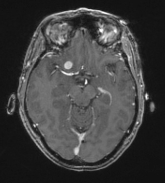
Washington University Experience | NEOPLASMS (MENINGIOMA) | Meningioma - Fibrous | 9A Meningioma, fibrous, intraventricular (Case 9) T1W - Copy
Case 9 History ---- The patient is a 59-year-old woman with past medical history of epilepsy, craniotomy for temporal lobe resection, migraine headaches, and deep vein thrombosis who is admitted for elective right frontal craniotomy for tumor resection. Per electronic medical record MRI shows a stable extra-axial homogeneously enhancing lesion arising from the right anterior clinoid process, most consistent with a meningioma. Operative procedure: Right anterior clinoid tumor resection. ---- 9A The small meningioma is seen in this T1-weighted contrast administered scan.

