Table of Contents
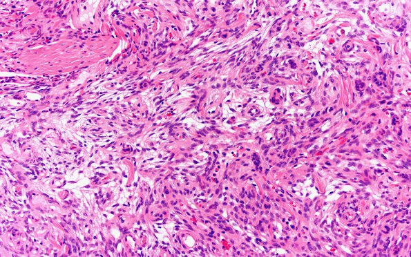
Washington University Experience | NEOPLASMS (MENINGIOMA) | Meningioma - Fibrous | 9B1 Meningioma, fibrous, intraventricular (Case 9) H&E 20X 2
9B1-4 H&E stained sections of the "anterior clinoid mass” show a well-circumscribed spindle cell neoplasm which has a predominantly cellular growth pattern with dense collagen bundles interspersed within the tumor. There are also hypocellular areas with a microcystic appearance. The tumor cells have round to ovoid nuclei with vesicular chromatin, intranuclear pseudoinclusions, and a modest amount of eosinophilic cytoplasm. Mitotic figures are rare. Atypical features, e.g. sheeting architecture, small cell change, macronucleoli, spontaneous necrosis, and hypercellularity, are not appreciated.

