Table of Contents
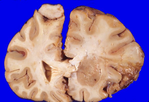
Washington University Experience | NEOPLASMS (MENINGIOMA) | Gross Pathology | 11A Meningioma (Case 11) gross 1
Case 11 History ---- This 79 year old female suddenly became unresponsive. Past history included hypertension, atherosclerotic coronary heart disease and atrial fibrillation. A CT scan showed a massive right internal carotid stroke, an old left-sided infarct and multiple small meningiomas located in the left frontal, suprasellar, and right CP angle. The patient was treated with Decadron and supportive care; however, she became increasingly obtunded with decerebrate posturing, fixed pupils and expired. ---- At autopsy there was right internal carotid thrombotic occlusion and acute right anterior cerebral and middle cerebral arterial infarcts with marked edema. She developed uncal herniation (right > left) with Duret brainstem hemorrhages. ---- There is a 2 cm. in diameter meningioma compressing the left superior frontal gyrus and damaging the underlying white and gray matter.

