Table of Contents
Washington University Experience | NEOPLASMS (MENINGIOMA) | Gross Pathology | 15C2 Meningioma, anaplastic (Case 15) H&E 11
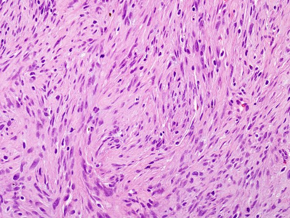
In a second pattern the tumor has a more fascicular spindled sarcomatous pattern. Mitotic figures are frequently present but relatively less brisk than those in the epithelioid appearing areas. ---- Immunohistochemical studies show patchy positivity of tumor cells for EMA in both these areas although weaker in spindled areas. A few clusters of tumor cells with epithelioid morphology are positive for progesterone receptor. Ki-67 labeling index is high reaching up to ~30% in more epithelioid looking areas and ~16.7% in more spindled areas. FISH revealed deletion of chromosome 1p and polysomy of chromosome 9 in epithelioid areas. In spindled areas, the tumor shows combined deletions of chromosomes 1p and 14q and monosomy of chromosome 9. These genetic alterations (deletions of 1p and 14q and monosomy of chromosome 9) are more often seen in higher grade meningiomas. ---- Comment: Although the tumor fails to reach a mitotic rate of >20/10HPF, often used as a separation point between atypical and anaplastic meningioma, the high grade histomorphological features (carcinomatous and sarcomatous areas), high mitotic rate (~17/10hpf) and high Ki-67 labeling index as well as genetic alterations (as evidenced by FISH), are consistent with anaplastic meningioma WHO Grade III and indicate an aggressive biological behavior.

