Table of Contents
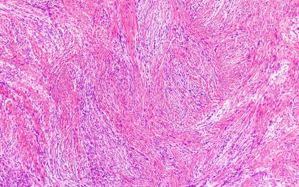
Washington University Experience | NEOPLASMS (MENINGIOMA) | Gross Pathology | 20C2 Meningioma, intraventricular (Case 20) H&E 10X
20C2-4 Higher magnification images show a predominantly fibroblastic meningioma arranged in a fascicular pattern with focal cellular whorl formation. The tumor cells have abundant eosinophilic cytoplasm, oval to spindled nuclei, finely granular to vesicular chromatin, and generally inconspicuous nucleoli. Mitotic figures are overall infrequent but focally up to 3 mitoses per 10HPF. Nodular hypercellularity is present. No necrosis or small cell changes are identified. (H&E)

