Table of Contents
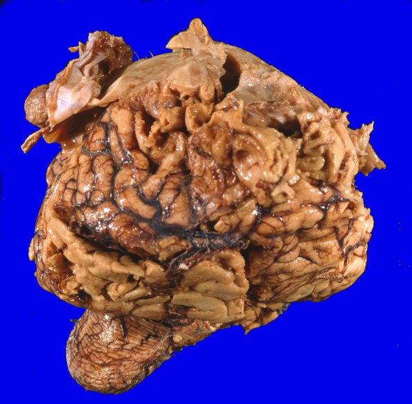
Washington University Experience | NEOPLASMS (MENINGIOMA) | Gross Pathology | 21A1 Meningioma, WHOII, brain invasion (Case 21) 2A
21A1,2 At autopsy the weight of the unfixed brain was 1430g and consisted of extracranial skin, soft tissue, bone, dura and brain. The scalp shows an 8 X 7 cm transcranial tumor with abscess. The abscess extends through soft tissue and bone overlying the right frontal lobe to show a 6 X 5 cm intracranial tumor that attaches to the dura, which was otherwise thickened over a 10 cm area. Multiple satellite nodules or distinct intracranial tumors are also attached to the dura, the largest of which measured 3 X 2.5 cm and surrounded the left posterior frontal lobe. The brain immediately adjacent to the tumor, including the parafalcine cortex, is markedly softened, friable and necrotic.

