Table of Contents
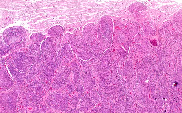
Washington University Experience | NEOPLASMS (MENINGIOMA) | Gross Pathology | 22C1 Meningioma (Case 22) 2X H&E N14 4X H&E
22C1,2 The tumor is a meningioma composed of epithelioid and spindled tumor cells with ovoid, hyperchromatic nuclei, frequent intranuclear pseudoinclusions, moderate amounts of eosinophilic cytoplasm, and often indistinct cell borders. In many areas, the tumor cells are arranged in fascicles and whorls; in others, the tumor is hypercellular, the architecture is vaguely sheeted, and there are many foci of small cell change. Scattered calcifications (some rounded, some elongated, representing mineralized collagen) are present. Mitotic figures are common. Macronucleoli and spontaneous necrosis were not appreciated. Brain invasion is also identified and, thus, the diagnosis of atypical meningioma with brain invasion, WHO grade II was made.

