Table of Contents
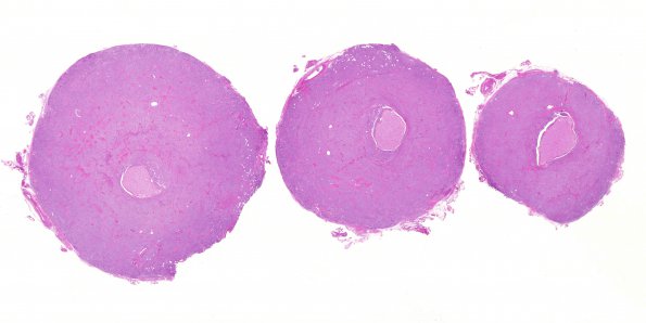
Washington University Experience | NEOPLASMS (MENINGIOMA) | Gross Pathology | 23C1 Meningioma, Optic nerve sheath (Case 23) H&E whole mount 2
23C1-3 Histological sections of the intraorbital mass show a neoplasm composed of tumor cells with round to oval nuclei, finely speckled chromatin, inconspicuous nucleoli, eosinophilic cytoplasm, indistinct cell borders, and occasional intranuclear clearings and pseudoinclusions. The tumor cells are organized into whorls and short, poorly formed fascicles by very thin collagen bundles, and into larger irregular lobules by thicker collagen bundles. Mitotic figures are rare, appearing at a density less than one per 10HPF. There is no evidence of 'sheeting' (loss of architecture), macronucleoli, necrosis, hypercellularity, or 'small cell change' (increased nucleus-to-cytoplasm ratio). Although the tumor surrounds the optic nerve circumferentially, there is no evidence of optic nerve invasion, or of tumor tissue at the posterior resection margin. ---- This histological pattern is that of meningioma, WHO grade I.

