Table of Contents
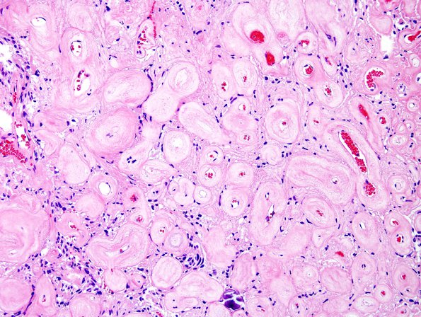
Washington University Experience | NEOPLASMS (MENINGIOMA) | Gross Pathology | 24C2 Meningioma, whorling sclerosing (Case 24) H&E 11
This is a meningothelial neoplasm with a range of morphologic features. There is the mild nuclear pleomorphism, with the majority of tumor cells containing round to oval nuclei and abundant eosinophilic cytoplasm. Many of the nuclei have clear holes or nuclear pseudo inclusions. Some areas show a papillary architecture with extensive non-calcifying collagenous whorls of varying size. There is marked vascular hyalinization. The tumor cells focally infiltrate into the adjacent brain parenchyma with extensive infiltration into the overlying dura and bone. Mitotic figures are not easily identified and there is no tumor necrosis.
Ancillary findings (not shown in this section of the atlas):
Immunohistochemical stains for progesterone receptor shows diffuse strong positive staining in tumor cell nuclei and membrane staining with EMA. The glial fibrillary acidic protein highlights invaded brain parenchyma. The Ki-67 proliferation index is low. The trichrome stain strikingly shows the hyalinized and fibrous areas. The histomorphologic and immunohistochemical stains are consistent with a whorling-sclerosing variant of meningioma with focal brain invasion, WHO grade II.

