Table of Contents
Washington University Experience | NEOPLASMS (MENINGIOMA) | Gross Pathology | 25A3 Meningioma (Case 25) 4
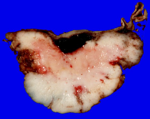
Gross appearance of this tumor as seen in cross section. ---- Microscopic description (not shown): The hypercellular, relatively solid appearing epithelioid neoplasm is arranged predominantly in sheets. Small cell formation is seen in several areas with clusters of cells with high nuclear/cytoplasmic ratio. Many tumor cells have macronucleoli, and foci of tumor necrosis are identified. Only rare mitotic figures are seen. A single section shows a small fragment of adjacent brain parenchyma; no invasion is identified. The tumor shows patchy membrane-pattern immunoreactivity for EMA and are largely negative for progesterone receptor. The Ki-67 labeling index is moderate, reaching 7.8%.---- Comment: The morphologic and immunohistochemical features are consistent with the diagnosis of atypical meningioma, WHO grade II.

