Table of Contents
Washington University Experience | NEOPLASMS (MENINGIOMA) | Gross Pathology | 2A3 Meningioma, suprasellar (Case 2) 5
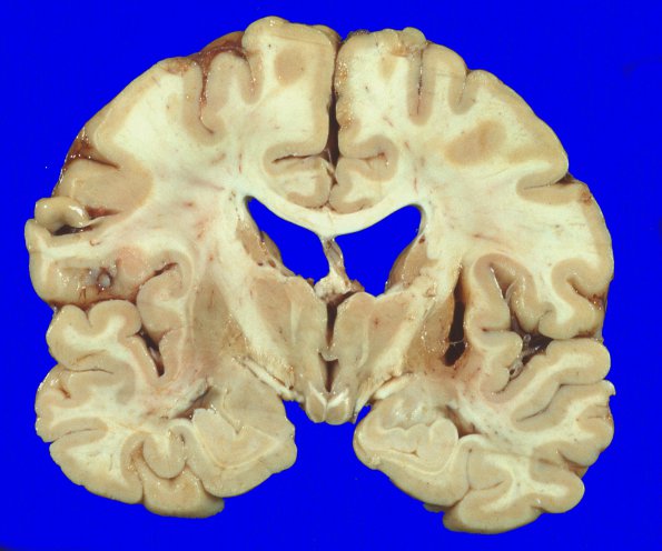
Coronal sections of the cerebral hemisphere showed an unremarkable cortical ribbon and underlying white matter. There was a 2 X 0.8 X 0.4 cm cystic cavity in the right putamen with yellow-brown discoloration, consistent with hemorrhage or hemorrhagic infarct. The right and left globus pallidus were small with orange discoloration and the substantia nigra pale and depigmented, both findings expected in NBIA1.

