Table of Contents
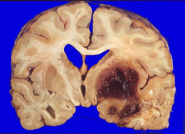
Washington University Experience | NEOPLASMS (MENINGIOMA) | Gross Pathology | 30A2 Meningioma, post surgical resection (Case 30) gross 1
Coronal sections through the cerebral hemispheres showed a large area of pale and hemorrhagic infarction involving the inferior aspects of the right frontal, temporal, and parietal lobes, including the right globus pallidus and putamen and the most inferior aspects of the internal capsule. Step sectioning of the vasculature at the base of the brain revealed 75% luminal occlusion but absence of thrombus or tumor invasion in the right middle cerebral artery. The right frontal and temporal lobes and basal ganglia show a resolving infarct consisting of confluent areas of neuronal necrosis and astrocytosis with numerous lipid-laden and hemosiderin-containing macrophages, capillary proliferation and, in the basal ganglia, large areas of confluent hemorrhage, consistent with occurrence at the time of surgery. Its distribution is consistent with interruption of penetrating vessels within the tumor bed.

