Table of Contents
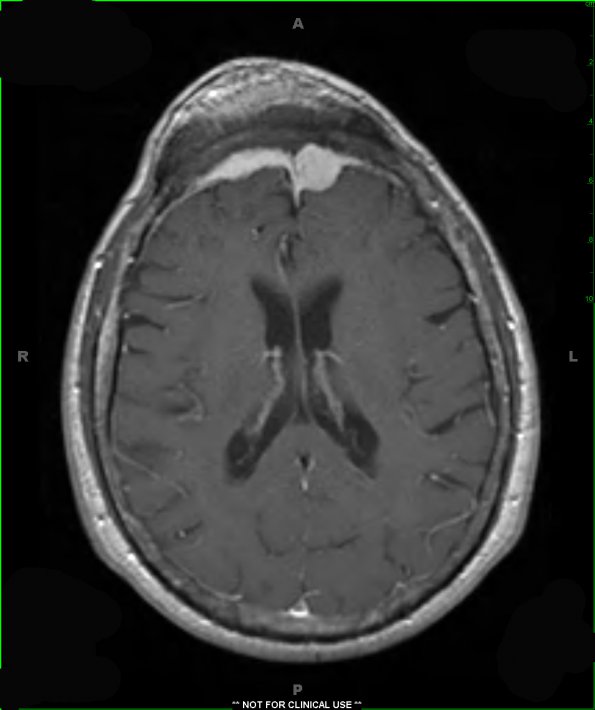
Washington University Experience | NEOPLASMS (MENINGIOMA) | Gross Pathology | 31A2 Meningioma & Hyperostosis (Case 31) T1 W 7 - Copy
A T1-weighted contrast applied scan demonstrates a lobular dural based mass within the left frontal lobe that appears to replace the marrow of the overlying frontal bones bilaterally and invade the soft tissues of the overlying scalp.

