Table of Contents
Washington University Experience | NEOPLASMS (MENINGIOMA) | Gross Pathology | 31B Meningioma & Hyperostosis (Case 31)_4
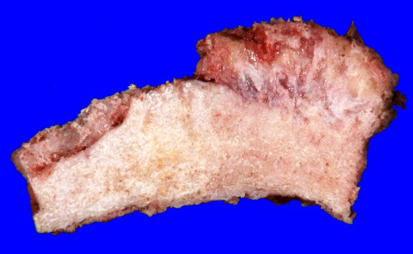
The neurosurgical specimen demonstrating hyperostosis resulting from tumor infiltration. Tumor tissue is present throughout the trabecular spaces of the expanded skull infiltrating soft tissue at the superficial border. ---- Findings described (not shown): Sections of the resected material show a collagen-rich hypercellular meningioma that exhibits several histologic patterns, including fibroblastic, meningothelial, sclerosing, and, focally, clear cell. Hypercellularity, focal 'small cell change', macronucleoli are described and mitoses reach 4 /10 HPF. The histological findings detailed above describe an atypical meningioma, WHO grade II; a grade II diagnosis is substantiated by a mitotic index of 4 (or more) per 10 hpf and, independently, by the presence of at least three of five contributing atypical features (in this case: hypercellularity, 'small cell change,' and macronucleoli).

