Table of Contents
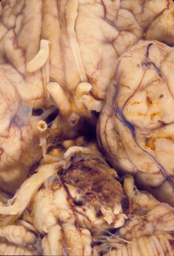
Washington University Experience | NEOPLASMS (MENINGIOMA) | Gross Pathology | 32A2 Meningioma removal, SP (Case 32) 3
There is a subarachnoid hemorrhagic area on the left temporal lobe surface, measuring I cm in greatest surface dimension. There are several hemorrhagic lesions involving the left temporal lobe. The hemorrhagic area in the left temporal lobe is a superficial hemorrhagic infarct with surrounding gliosis, vascular proliferation and hemosiderin laden macrophages.

