Table of Contents
Washington University Experience | NEOPLASMS (MENINGIOMA) | Gross Pathology | 37A1 Meningioma (Case 37) - Copy
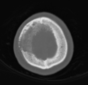
Case 37 History ---- The patient was a 71 year old woman with mild weakness of the left upper and lower extremities. CT with contrast showed a large extra-axial hyperdense lesion in the right frontal lobe that invaded the skull and exerts mass effect on the ventricle. Operative procedure: Craniotomy for tumor resection and replacement of skull defect. ---- 37A1-3 Radiographic studies: 37A1 In this case CT scan shows a cellular neoplasm involving the dura mater and overlying bone. Within the bone, the trabecular spaces are enlarged due to the infiltration of tumor and there is remodeling of the bone with many thinned areas.

