Table of Contents
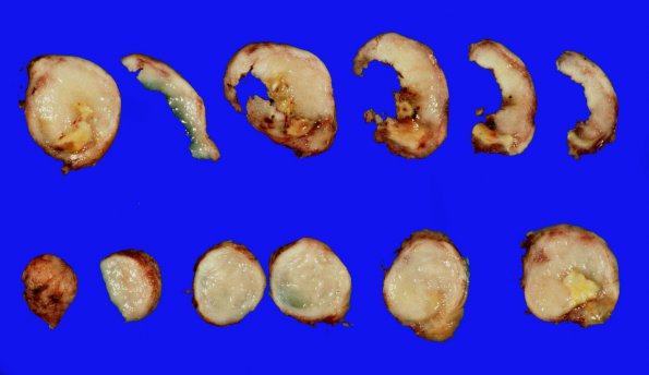
Washington University Experience | NEOPLASMS (MENINGIOMA) | Gross Pathology | 38A2 Meningioma (Case 38) 3
Cross sections show patchy necrotic foci. ---- A hypercellular neoplasm that is composed predominantly of large nests of epithelioid cells surrounded by variable quantities of collagen septae. Prominent whorl formation and occasional psammoma bodies are seen. In some areas, the tumor is composed of fibroblast-like spindle cell tumors that form intersecting fascicles with prominent collagen deposition. The tumor cells show oval to elongated nuclei with pseudoinclusions and abundant cytoplasm. The tumor invades the dura, and extensive necrosis is seen in multiple sections of the tumor. However, the tumor does not appear to be mitotically active and shows no sheeting architecture, small-cell formation or macronucleoli. No brain invasion is identified, although a few fragments of brain tissue abut the tumor. Overall, the morphologic features and immunophenotype are diagnostic of meningioma, WHO grade I, with increased proliferation indices.

