Table of Contents
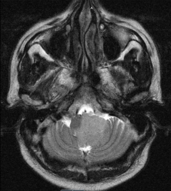
Washington University Experience | NEOPLASMS (MENINGIOMA) | Gross Pathology | 3A1 Meningioma (Case 3) T2 - Copy
Cerebral angiogram and MRI showed a 2.7 X 3.6 X 3.4 cm extra-axial mass in the left posterior fossa that compressed the medulla, encased the distal left vertebral artery and narrowed the basilar artery.

