Table of Contents
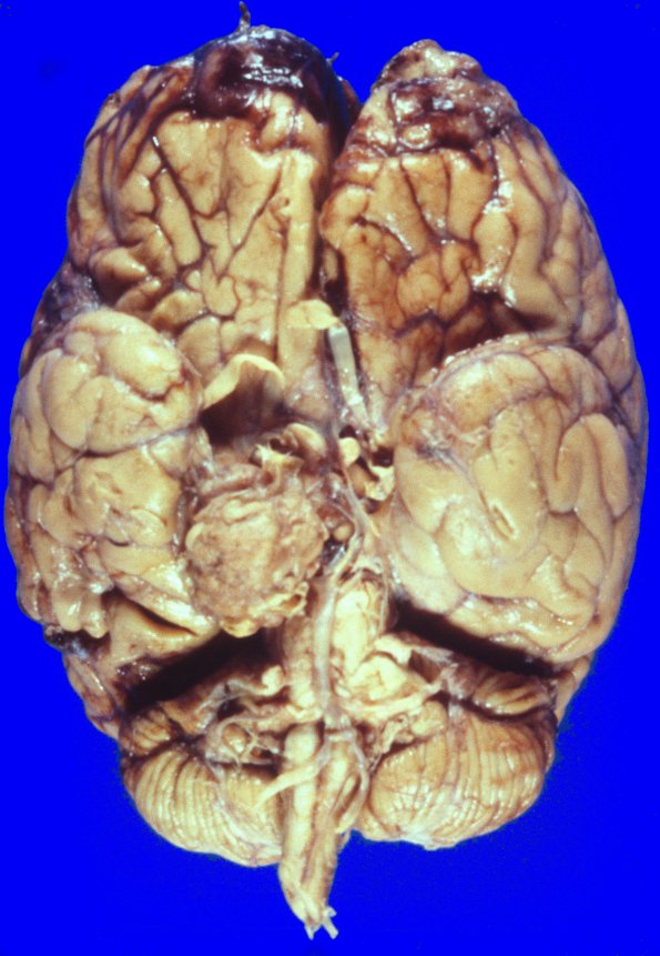
Washington University Experience | NEOPLASMS (MENINGIOMA) | Gross Pathology | 51A1 meningioma, parasellar (Case 51)
The examination of the basal aspect of the brain shows a 3.5 cm mass attached to the leptomeninges of the medial aspect of the right temporal lobe, compressing it with extension into the right orbit. The medial aspect of the right temporal lobe and the right uncus are compressed by the mass. The right optic nerve and right lateral aspect of the optic chiasm are focally indented by the mass.

