Table of Contents
Washington University Experience | NEOPLASMS (MENINGIOMA) | Gross Pathology | 57A1 Meningioma (Case 57) 1
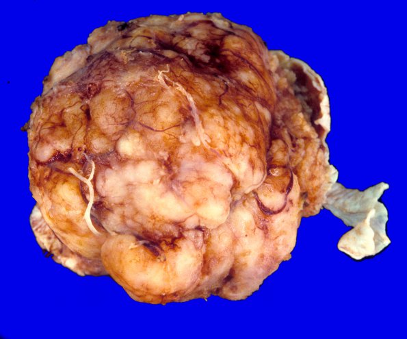
Case 57 History ---- The patient was a 78 year old right-handed man admitted to the medicine service for loss of consciousness and urinary incontinence, followed by confusion that lasted 30-60 minutes. An MRI showed a 3 X 2 cm enhancing lesion in the left frontal region anteriorly. Clinical diagnosis: Brain tumor. Operative procedure: Craniotomy for excision of tumor. ---- 57A1,2 The gross neurosurgical specimen in external and cut section. ---- Sections show a meningioma with atypical histologic features. The neoplasm is composed of spindled cells with eosinophilic cytoplasm that demonstrate prominent "whorling" architecture. One area of the neoplasm is relatively hypercellular and in this region there is an increased mitotic rate, although the mitotic rate in the rest of the neoplasm appears low. The neoplasm shows small areas of necrosis throughout and some cells have large enlarged, hyperchromatic nuclei. No brain tissue is associated with the neoplasm. ---- This is an old case and was called atypical meningioma, although it would not qualify for that diagnosis using modern criteria.

