Table of Contents
Washington University Experience | NEOPLASMS (MENINGIOMA) | Gross Pathology | 60A2 Meningioma (Case 60) Gross _2
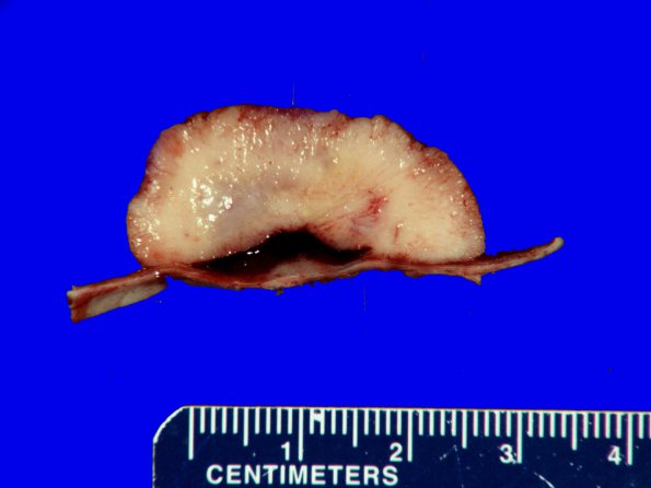
A typical appearance of a bossellated tumor seen in cross section. ---- A sheeting growth pattern, small cell change, necrosis and prominent nucleoli, is consistent with an atypical meningioma designation (WHO grade II). The Ki-67 proliferation index is elevated focally reaching ~13.3%. ---- Patchy areas of brain invasion, supported by a GFAP immunostain, were also seen. By immunohistochemistry, the neoplastic cells show multiple, minute, foci of epithelial membrane antigen expression. The tumor lacked nuclear expression of progesterone receptor in most parts and showed only rare positivity. The overall findings are those of an atypical meningioma with patchy, superficial, brain invasion (WHO grade II). The designation of WHO grade II is given based upon the brain invasion as well as presence of atypical features including hypercellularity, necrosis, sheeted growth pattern, small-cells, and prominent nucleoli. In addition, markedly elevated Ki-67 proliferation index in this meningioma is a worrisome finding and this may portend an aggressive outcome.

