Table of Contents
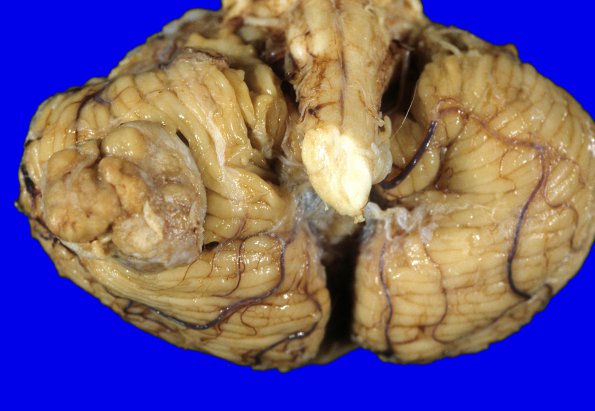
Washington University Experience | NEOPLASMS (MENINGIOMA) | Gross Pathology | 61A1 Meningioma, cerebellum (Case 61) 2
Case 61 History ---- The patient was a 78-year-old woman with a long history of Parkinsonism, depression with psychotic features, degenerative joint disease, and hypothyroidism. MRI imaging of the patient's head in March, 1999, showed a 2 cm right cerebellar enhancing mass of concern for a metastatic lesion; however, work-up revealed no primary malignancy. Cerebrospinal fluid cytology showed atypical lymphoid cells, highly suspicious for malignant lymphoma; however, flow cytometry failed to show diagnostic features of a neoplastic process. The mass was not visualized in a 2002 computed tomography study of the patient's head. The patient died shortly thereafter without cerebellar symptoms. ---- At autopsy, the weight of the unfixed brain is 990 g. The dura in the base of the right posterior fossa shows a 2.5 cm, firm, tan-white mass that indents, but does not appear to invade the cerebellum. Radial sections of the cerebellum show normal foliar architecture generally but distortion of the cortex in the region of the superficial mass. The cortex, white matter, and dentate nucleus are unremarkable. ---- The mass involves the inferior aspect of the right cerebellum

